Project Description
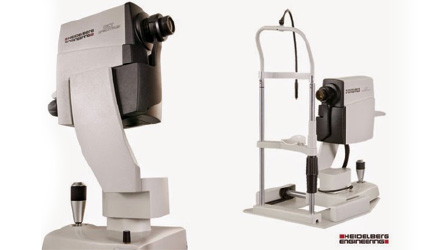
Heidelberg Spectralis OCT
(Ocular Coherence Tomography)
The Spectralis OCT allows us to obtain microscopic images of the retina (back of the eye) to aid in the diagnosis and treatment or many diseases such as glaucoma, diabetic eye disease, and age-related macular degeneration.
The OCT uses a principle similar to ultrasound, yet, instead of sound waves, uses laser light to obtain an image. The OCT is 10 times more sensitive than ultrasound, with the ability to display objects as small as one hundredth of a millimetre wide.
We have just purchased the newest model of the Spectralis OCT which allows us to offer simultaneous dual-beam scanning for multi-modality imaging consisting of: Spectral-Domain OCT (SD-OCT), Fluorescein Angiography, ICG Angiography, Autofluorescence, Infrared Imaging and Red-Free Imaging.
We are able to diagnose and monitor a number of conditions with the Spectralis OCT such as Age Related Macular Degeneration, Diabetic Retinopathy, Retinal Vein Occlusions and Glaucoma.

